Boon’s Broken Tooth
Published: July 31st, 2021
Boon’s Broken Tooth
Published: July 31st, 2021
THIS ARTICLE IS FROM THE Summer/Autumn 2021 EDITION OF OUR Pets & Vets Magazine
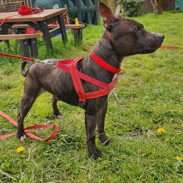
This is Boon, a lively 3 year old Staffie. He was found and brought to us here at Stray Aid as his owner couldn’t be found. Unfortunately, as Boon was not microchipped, not wearing a collar with his owner’s details, and no one came forward for him, we were unable to trace his family. As we couldn’t access his medical history, we gave Boon new vaccinations, flea and worm treatments, neutered him, and gave him a new microchip to stop him from getting lost in the future. During his initial examination by our vet, we found that Boon had somehow broken one of his upper canine teeth before he arrived at Stray Aid.
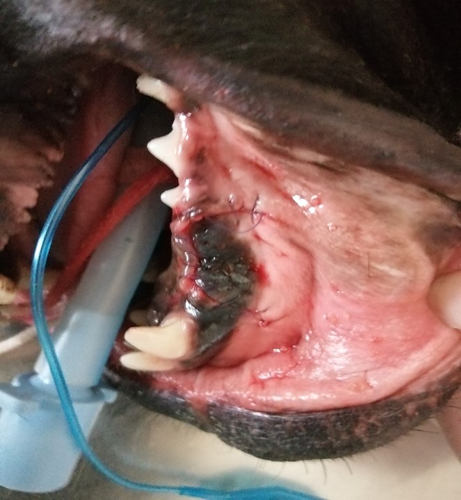
The tooth had recently broken, and the break extended below the gumline. The sensitive pulp cavity (inner part of the tooth, containing nerves) was exposed, causing pain for Boon. To relieve his pain, the tooth was carefully and painstakingly extracted under quite a long period of general anaesthesia.
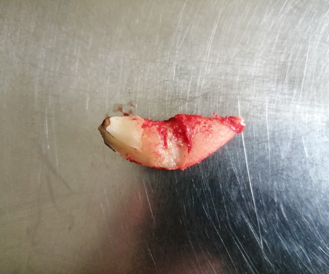
As you can see from the photograph, the root (the portion on the right, with blood) of a canine tooth is much larger than the crown of the tooth (the part covered in enamel and which is visible from the gumline) so the crown of the tooth is “the tip of the iceberg”.
In dogs, the majority of tooth extractions are a result of periodontal disease, whereby the accumulation of plaque and tartar causes the gums to recede and the supporting ligaments around the tooth to be broken down. These teeth become loose and are usually not too difficult to remove because they are not held tightly and firmly into their sockets. Broken teeth are strongly held in place by tough ligaments which must be broken down before the teeth can be removed. The canine root is not only long, but curves into the jaw. The canine teeth in wild animals are used for gripping their prey, so the shape and structure means that they do not come out easily.
To extract Boon’s broken tooth, our vet cut a flap of tissue in the surface of his gum, pushed the flap back and then drilled away the bone covering the surface of the tooth root. She then broke down the ligaments on the surface and the tip of the root, before removing the broken tooth. After the tooth extraction, the flap of tissue was sutured back into place.
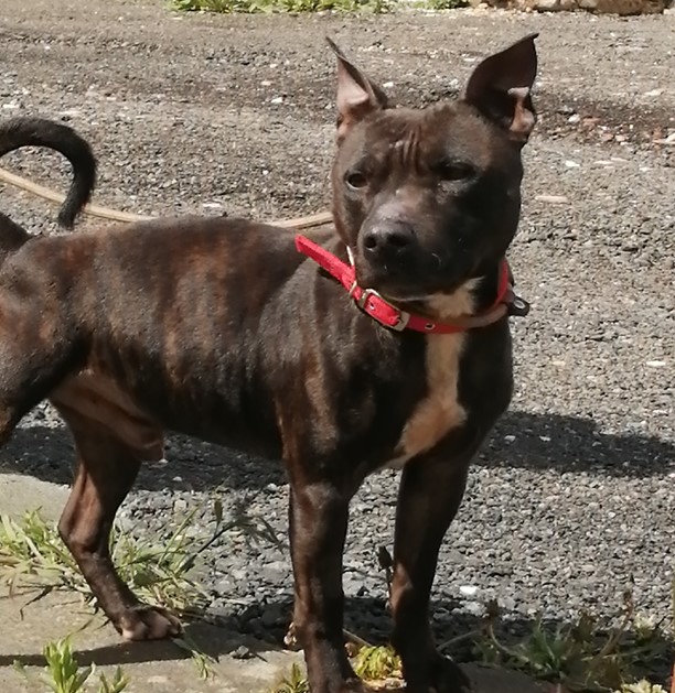
Boon also had a small skin tag removed, taking the opportunity whilst he was under general anaesthesia. He was given antibiotics and pain relief after the procedure, and soft food only for a week. Boon was given another thorough check over by our vet after a few days, and he was found to be happy, eating well and pain-free, and his gum had healed well at this stage.
We are delighted to say Boon has now gone to his loving new home where we wish him all the best for the future.
Support our vets today – join Team SAFE and help sponsor the care of the hundreds of dogs and cats we help each year